Forensic investigation on aquatic wildlife in Hong Kong and Greater China with Artec Ray & Leo
Challenge: To capture aquatic mammals as large as whales in full-color 3D, for forensic investigation, research, and conservation.
Solution: Artec Eva, Artec Space Spider, Artec Leo, Artec Ray, Artec Studio, CT scans
Result: Using an array of Artec 3D scanners, whales, dolphins, sea turtles, and more are captured, along with the boats and damaging surfaces that may have caused injury or death. Collected research findings and recommendations are shared with government bodies and stakeholders in the field such as shipping companies, with a goal of aiding precise conservation measures and finding effective solutions for injury prevention.
Why Artec 3D? With a wide range of scanners that capture everything from ventral grooves to the entire length and girth of a whale to whole ships and boats, Artec Studio steps up to combine all data for a complete 3D picture of these marine animals, contributing directly to the research, and to the essential conversation and conservations that follow.
When it comes to forensics and autopsies, most of us might imagine human bodies, hospitals, mortuaries, and labs. But in the scientific world, these terms extend much further – and they include our fellow mammals, though those of the sea-dwelling and much larger variety. And for one team in Hong Kong, 3D technology and forensic investigation of aquatic animals combine: 3D scanning everything from animal injuries, and the vessels that caused the damage.

A deceased dolphin being scanned with Artec Leo
“Our main research subjects are aquatic animals stranded in Hong Kong, and adjacent waters – animals who utilize the habitat range of the coastal waters along the South China Sea,” said Prof. Brian Kot, Assistant Professor and Veterinary Imaging Researcher in the Aquatic Animal Virtopsy Lab, City University of Hong Kong.
The aim of this research is if nothing else ambitious: to provide scientific and precise evidence of natural and anthropogenic impacts on aquatic animals through a virtopsy-led postmortem investigation approach – 3D documentation of stranded carcasses and suspected injury-inflicting tools and postmortem CT/MRI/ultrasound examination of the carcasses. This yields their complete e-copies for death investigation, matching analysis, and virtual accident scene reconstruction. In short, to document injured or deceased aquatic animals, to investigate how and why it happened, and to consider how to prevent such incidents in the future.

A dolphin being scanned with Artec Space Spider
“Vessel interaction was one of the major anthropogenic threats to the local resident cetaceans,” explained Prof. Kot. “In 2018, evidence of sharp- or blunt-force trauma indicative of vessel interaction was observed in 19 out of 38 stranded cetaceans we encountered.” While less maritime traffic following the COVID-19 pandemic in 2020 decreased such interactions, there are more threats still. In 2023, signs of fishing gear entanglement or peracute underwater entrapment were detected in 15 out of 25 stranded cetaceans that were reported.

The shell and body of a sea turtle being scanned
“We aim to advance our investigation towards the goal of marine conservation, and also dolphin or sea turtle conservation to facilitate science-based discussion and policy decisions – this allows the use of aquatic animals as sentinels of ecosystem health, working towards a ‘One Ocean-One Health’ ideal,” summarized Mr. Henry Tsui, the Senior Research Assistant and Manager in the Aquatic Animal Virtopsy Lab, City University of Hong Kong. “And in the event that there is a court case, these findings could act as evidence that is far more accurate and reliable than photographic 2D images.”
Growing the fleet
While the team first used conventional methods such as photography and photogrammetry to capture the external condition of the animals, these options fell short.
“Photography itself is affected by multiple parameters, including the lighting of the environment, distance, and a lot of things,” said Prof. Kot. “So we needed 3D models in order to visualize these skin conditions, wounds and injuries, and maybe the inflicting tools as well.”
Having tried various 3D scanning alternatives, the team eventually found a solution in Artec 3D.

A sea turtle as it was found, now captured in high-resolution 3D
“We picked Artec 3D firstly because color is extremely important to us. When we are investigating injuries and wounds, the texture actually gives us a lot more information: whether or not the wounds are new wounds, healing wounds, or is there any information, and so on.”
Starting off with Artec Eva, a versatile solution for capturing medium-sized objects, and Space Spider, an industry favorite in capturing fine details in super high accuracy, the team soon decided to expand their toolset. “We used Eva for scanning the entire animal body, and Space Spider for focal regions of interest like the head, dorsal fin, genital slit, and any wounds or skin lesions. That usually takes about 20-30 minutes for data acquisition,” Prof. Kot said. “We also use Artec Leo, and it takes just about 5-10 minutes for scanning a dolphin or porpoise.”

Getting up close with a whale using the wireless Artec Leo
Besides dolphins and porpoises, the team scans sea turtles. Wireless, AI-powered, and incredibly easy to use, Artec Leo was built for cases like this: Moving around a large surface, capturing quickly and without any wires or cables to get in the way.
Essential documentation
Because the goal is to scan and inspect not only the animal’s exterior but the interior too, scanning has to be done carefully, and very particularly.
“Our main goal is to combine both external and internal data together in order to have a holistic 3D model of the whole body, right?” explained Prof. Kot. “So, we put the animals on top of the CT scanning table, and what we do is we scan it in a systematic manner: the lateral side, that side view, and also the top view and the bottom.”

For a full data set, animals are scanned in CT and 3D
Following a thorough scan of the exterior of the carcass, the animal is then opened up for internal scanning. Due to the shifting position of the animal, a combination of external 3D scans as well as internal volumetric CT data is used to create complete models and carry out analysis using advanced rendering software such as Blender.
The full-body exhaustive documentation has proved innumerably helpful for the team, with a lot to be said for permanently available and thoroughly detailed evidence. ”By the time we open up the body, we can’t sew it up anymore, right, and that’s after the necropsy, after dissection – there is no way back,” said Prof. Kot. “So, having 3D data and models allows us to do multiple rounds of investigation – not just locally, but also internationally as well, as it could be shared among different groups of experts worldwide for further investigation as well.”

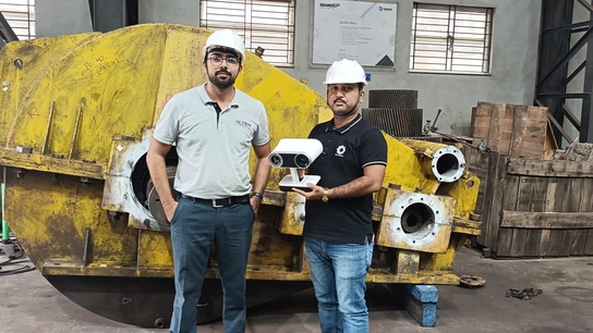
Artec Ray is used for capturing boats, ships, and surfaces that may have led to the animal’s injury or death
How to scan a whale in 10 mins
When dealing with items as large as whales and boats, it’s Artec Ray’s time to shine. “When we have large whales, it will create such a huge data set, so we use Ray to document them,” said Dr. Tabris Chung, Research Fellow and Scientific Coordinator in the Aquatic Animal Virtopsy Lab, City University of Hong Kong. With photogrammetry, the team can then map color into the 3D model, and obtain a highly detailed, accurate scan – even when working with the world’s largest mammal.
Data can also be used in collaboration with other investigations and projects that the team is working on. For example, addressing vessel collision as a threat to the dolphins that reside in these waters.

Tragically, these sea creatures are often already dead when found. Here, an entire whale is captured with Artec Leo
“We are able to find out where the hotspots of these accidents are, and also which types of vessels will appear around the waters,” Dr. Chung said. “Then we go to the shipyard, we go to the dockyard, and look at all these types of marine vessels. With Artec Ray, we scan the bottom of these marine vessels.”
With all the data successfully collected in high-resolution 3D, analysis and scene reconstruction are carried out.

Boat and ship parts are also collected as evidence
“Most importantly, this is able to help people understand how the marine vessels collide with these vulnerable dolphins, but at the same time, it allows people to understand how the injury could be prevented,” Dr. Chung said. “These are important, and the reasons behind why we use our Artec 3D scanners on almost everything we receive.”
Sharing their tools
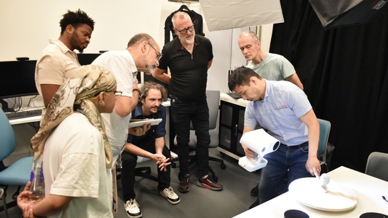
While the team remains hard at work collecting, presenting, and analyzing these aquatic animals, they’re also focused on the future. So far, they have hosted a 3D scanning segment as part of a post-mortem investigation workshop annually, in order to provide local, regional, and global participants with lessons on how 3D scanning and CT scans are used for postmortem investigation.
But as with all things, it is important to not only focus on the cause of death but on the environment and overall well-being of these magnificent sea creatures.

Workshops are held to demonstrate the use and power of 3D scanning
“Very importantly, we must consider the health concerns of the animals holistically rather than solely the cause of death,” said Prof Kot. “Infectious diseases are common in cetaceans, which could impair mobility due to verminous pneumonia or musculoskeletal diseases. Vision or hearing deficits can also result in reduced foraging capacity, and poorer response to the surroundings and greater risk of vessel interaction.”
“With the goal of setting up precise conservation measures for our endangered aquatic wildlife, we cannot simply blame the boats,” advised Prof. Kot, “but rather, we should pay more effort in understanding the biological health of these animals and try to mitigate the risks related to natural and anthropogenic impacts.”
Scanners behind the story
Try out the world's leading handheld 3D scanners.


